维管形成层(环)形成后,主要是进行切向平周分裂。向内分裂产生的细胞形成新的本质部,加在初生木质部的外方,称为次生木质部;向外分裂产生的细胞形成新的韧皮部,加在初生韧皮部的内方,称为次生韧皮部。次生木质邵和次生韧皮部,合称为次生维管组织,是次生结构的主要部分。在具有次生生长的根中,次生木质部和次生韧皮部间始终存在维管形成层。次生水质部和次生韧皮邵的组成,基本上和初生结构中相似,但次生韧皮部中,韧皮薄壁组织较为发达,韧皮纤维的含量较少。另外,在次生木质邰和次生韧皮部内,还有一些径向排列的薄壁细胞群,称为木射线( xylem ray )和韧皮射线( phloem ray),统称为维管射线( vascular ray)(图2-15)。维管射线是次生结构中新产生的组织,它从形成层处向内外贯穿次生木质部和次生韧皮部,作为横向运输的通道和结构。次生木质部导管中的水分和无机盐,就可以通过维管射线运送到形成层和次生韧皮部。同样,次生韧皮部中的有机养分,也叮以通过维管射线运送到形成层及次生术质部。维管射线的形成,使根的维管组织内有轴向系统(导管,管胞、筛管、伴胞,纤维等)和径向系统(射线)之分。
白色的线为维管射线。
根的次生生长
In secondary growth, primary tissues and residual meristematic tissues produce secondary meristems, which then produce secondary tissues. Whereas primary tissues allow for vertical growth, secondary tissues allow for lateral growth: they allow stems and roots to become wider by producing wood. In addition to growing wider, secondary growth exchanges the living epidermis for a thick layer of dead, waterproofed cells called cork. The cork and a few other layers of tissue comprise something called the periderm, or perhaps more familiarly called bark.
This type of growth first evolved in gymnosperms. It is also present in many angiosperms. As a general rule, monocots do not undergo secondary growth (though some, like bamboo, have an analogous相似的 process).
Gymnosperms裸子植物
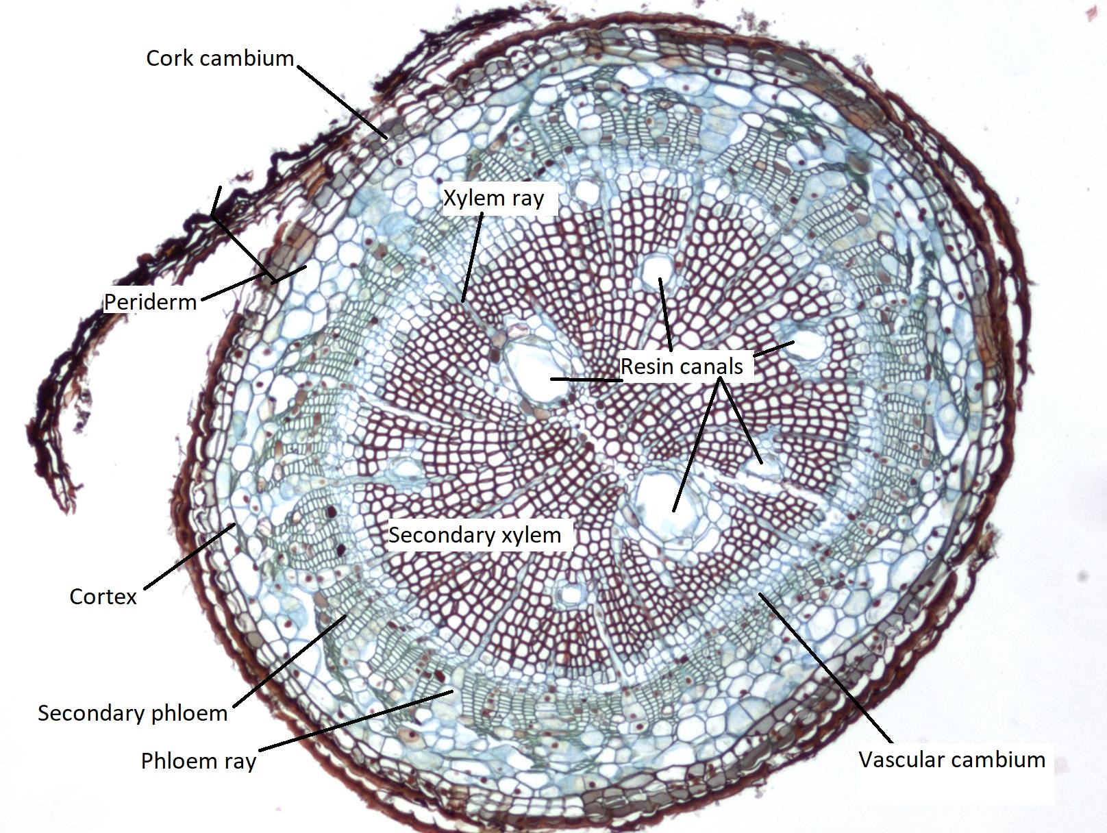
Figure \(\PageIndex{1}\): A cross section of a pine root in the early stages of primary growth. The first layer of periderm is forming, causing the epidermis to slough off. The periderm is currently the outermost layer, compose of a few layers of suberized cork cells, the cork cambium, and large cells called the phelloderm. The cortex is just inside the phelloderm and is getting smaller as the root grows. Ranks of cells just inside the cortex are secondary phloem cells (stained a blue-green color) which are being produced by the vascular cambium (a ring of cells stained light blue between the xylem and phloem). Just within the vascular cambium, making up the center of the root, is the secondary xylem (stained red, due to the secondary wall). There are large holes in the secondary xylem where resin canals are traveling through. Strings of parenchyma cells traverse laterally through the vascular tissue. These are called xylem rays and phloem rays, depending on their location within the vascular tissue. Image from Berkshire Community College Bioscience Image Library, CC0, via Wikimedia Commons.
Angiosperms被子植物
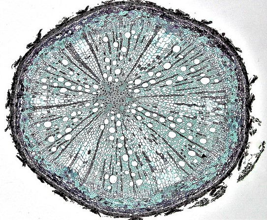
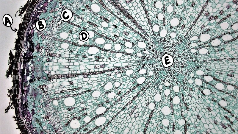
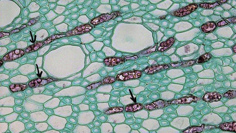
Figure (): Older Quercus root, 40x. "In young roots, the vascular cylinder is surrounded by two rings of cells, the pericycle and endodermis, and above these layers the cortex and epidermis. In older roots underling activity of the cork cambium replaces the epidermal and cortical tissues with a protective zone of cork rich periderm. The outermost layer of periderm consists of layers of cork cells, the phellem, which produce the waterproofing substance suberin. Cork cells are dead at maturity. Deep to the phellem is a layer of living cork cambium or phellogen and just beneath that layers of cork parenchyma or phelloderm. Many cells in the periderm contain dark staining tannins. The vascular cylinder consists of an outer narrow ring of phloem,and deep to this, the vascular cambium. The vascular cambium remains active, producing annual growth of secondary phloem towards the outside of the root and secondary xylem towards center of the root. Because of greater production of xylem, the bulk of the vascular cylinder is dominated by radially arranged rays of secondary xylem interspaced with medullary rays of parenchyma cells. Annual growth rings of spring and summer wood is difficult to distinguish in roots. Both the pericycle and endodermis, which wrap vascular cylinder in younger roots, are lost due to seasonal growth of the vascular cylinder. The center of the root is made up of primary xylem." Caption text and image from Berkshire Community College Bioscience Image Library, CC0, via Wikimedia Commons.Figure (): A labeled cross section through an older Quercus root, 100x. A=Periderms, B=Secondary Phloem, C=Vascular Cambium, D=Secondary Xylem, E=Primary xylem. Image from Berkshire Community College Bioscience Image Library, CC0, via Wikimedia Commons. Labels added by Maria Morrow.Figure \(\PageIndex{4}\): A labeled cross section through the outer tissues of an older Quercus root, 400x. A=Cork cells, B=Cork cambium, C=Phelloderm (A, B, and C=Periderm), D=Secondary phloem fibers, E=Secondary phloem, F=Vascular cambium, G=Secondary xylem. Image from Berkshire Community College Bioscience Image Library, CC0, via Wikimedia Commons. Labels added by Maria Morrow.Figure \(\PageIndex{5}\): A cross section through the secondary xylem of an older Quercus root, 400x. The rows of darker cells (indicated by arrows) are parenchyma cells that traverse the secondary xylem and phloem. In the xylem, they are called xylem rays. In the phloem, they are called phloem rays. Image from Berkshire Community College Bioscience Image Library, CC0, via Wikimedia Commons. Labels added by Maria Morrow.
 就较多,特别是内方产生的新组织(次生木质部)也较多,这样就把形成层环向外较大的推移,结果使得性个形成层环从横切面上看,成为较为整齐的圆形,此后,形成层的分裂活动也就按照等速进行,有规律地形成新的次生结构,并将初生韧皮部推向外力。